2 New Research Awards (January 2023)

January 30, 2024

From the UNC/NC State Joint BME News announcements: https://bme.unc.edu/2024/01/dr-jason-franz-receives-two-awards-to-accelerate-wearable-sensing-to-optimize-knee-joint-health/

BME Associate Professor Dr. Jason Franz has established a highly productive and collaborative line of research that integrates wearable sensing and machine learning for precision rehabilitation of individuals with knee osteoarthritis. That research, in close partnership with Dr. Brian Pietrosimone from the UNC Department of Exercise and Sports Science, was recently recognized with two awards to accelerate their path from scientific discovery to commercialization and genuine translational impact.

The first, a 2-year $110k translational research grant from the North Carolina Biotechnology Center, will generate patient data to demonstrate proof-of-concept and feasibility of a novel wearable sensing and machine learning prediction technology for detecting, treating and monitoring aberrant forces during walking relevant to the onset and progression of knee osteoarthritis.

The second, a $50k commercialization grant from UNC Kickstart Venture Services, was awarded to VETTA Solutions – the start-up company inspired by these research discoveries and co-founded by Drs. Franz and Pietrosimone.

Commercialization and entrepreneurship are cornerstones of our mission here in BME, and we want to congratulate Dr. Franz and his entire team for their recent success.”

Paper Accepted (November 2023)

November 3, 2023

The effect of prolonged walking on leg muscle activity patterns and vulnerability to perturbations. Journal of Electromyography and Kinesiology.

Abstract:
Understanding the consequences and ecological relevance of muscle fatigue is important to guide the development of strategies to preserve independence. However, few studies have examined walking-related fatigue and the effects on walking instability. Our purpose was to investigate the effects of prolonged walking on leg muscle activity and vulnerability to balance perturbations. Eighteen healthy young adults completed a 30-min walking trial at their preferred walking speed while leg muscle activities were recorded. Before and after the 30-min walk, participants responded to five 5% body weight lateral force perturbations. Time-frequency analysis with wavelet transformation and principal component analyses assessed neuromuscular adaptations of muscles to prolonged walking. Following prolonged walking, we observed a time-dependent increase in EMG intensities at slower frequencies for the soleus and tibialis anterior and a decrease in mean amplitudes for the soleus, lateral gastrocnemius, and semitendinosus. Mean mediolateral CoM displacement following perturbations averaged 21% larger after the 30-min walk. Our results suggest that walking for 30 minutes at a comfortable speed elicits complex neuromuscular adaptations indicative of local muscle fatigue and an increased vulnerability to walking balance perturbations. These findings could inform fatigue monitoring systems or walking assistive devices aimed at reducing walking-related fatigue and maintaining independent mobility.

Paper Accepted (November 2023)

November 3, 2023

Does the effect of walking balance perturbations generalize across contexts? Human Movement Science.

Abstract: Balance perturbations are used to study locomotor instability. However, these perturbations are designed to provoke a specific context of instability that may or may not generalize to a broader understanding of falls risk. The purpose of this study was to determine if the effect of balance perturbations on instability generalizes across contexts. 29 healthy younger adults and 28 older adults completed four experimental trials, including unperturbed walking and walking while responding to three perturbation contexts: mediolateral optical flow, treadmill-induced slips, and lateral waist-pulls. We quantified the effect of perturbations as an absolute change in margin of stability from unperturbed walking. We found significant changes in mediolateral and anteroposterior margin of stability for all perturbations compared to unperturbed walking in both cohorts (p<0.042). However, in older adults only mediolateral effects of waist-pull perturbations and optical flow perturbations and waist-pull perturbations and treadmill-induced slips correlated significantly (r ≥0.398, p-values≤0.036). In younger adults but not in older adults, we found positive and significant correlations between the anteroposterior effect of waist-pull perturbations and optical flow perturbations, and the anteroposterior and mediolateral effect of treadmill-induced slips (r≥0.428, p-values≤0.021). We found no “goldilocks” perturbation paradigm to endorse that would support universal interpretations about locomotor instability. Building the most accurate patient profiles of instability likely requires a series of perturbation paradigms designed to emulate the variety of environmental contexts in which falls may occur.

Paper Accepted (October 2023)

October 26, 2023

Sustained limb-level loading: A ground reaction force phenotype common to individuals at high-risk for and those with knee osteoarthritis. Arthritis & Rheumatology.

OBJECTIVE: To compare the vertical (vGRF), anterior-posterior (apGRF) and mediolateral (mlGRF) ground reaction force (GRF) profiles throughout the stance phase of gait: 1) between individuals 6-12 months post anterior cruciate ligament reconstruction (ACLR) and uninjured matched controls; and 2) between ACLR and individuals with differing radiographic severities of knee osteoarthritis (KOA) defined as Kellgren and Lawrence (KL) grades KL2, KL3, and KL4.METHOD: A total of 196 participants were included in this retrospective cross-sectional analysis. Gait biomechanics were collected from individuals 6-12 months post-ACLR (n=36), uninjured controls matched to the ACLR group (n=36), and individuals with KL2 (n=31), KL3 (n=67), and KL4 OA (n=26). Between-group differences in vGRF, apGRF, and mlGRF were assessed in reference to the ACLR group throughout each % of stance phase using a functional linear model.RESULTS: The ACLR group demonstrated lesser vGRF and apGRF in early and late stance compared to the uninjured controls, with large effects (d range: 1.35-1.66).
Conversely, the ACLR group exhibited greater vGRF (87-90%; 4.88%BW; d=0.75) and apGRF (84-94%; 2.41%BW; d=0.79) than the KL2 group in a small portion of late stance. No differences in mlGRF profiles were observed between the ACLR and either the uninjured controls or the KL2 group. The magnitude of difference in GRF profiles between the ACLR and OA groups increased with OA disease severity.CONCLUSION: Individuals 6-12 months post-ACLR exhibit strikingly similar GRF profiles as individuals with KL2 KOA, suggesting both patient groups may benefit from targeted interventions to address aberrant GRF profiles.

Paper Accepted (October 2023)

October 11, 2023

Ostovari, Yu research published in PLOS ONE - Healthcare Ergonomics Analytics Lab - Purdue UniversitySimulations suggest walking with reduced propulsive force would not mitigate the energetic consequences of lower tendon stiffness. Ricky Pimentel, Greg Sawicki, and Jason R. Franz

Abstract. Aging elicits numerous effects that impact both musculoskeletal structure and walking function. Tendon stiffness (kT) and push-off propulsive force (FP) both impact the metabolic cost of walking and are diminished by age, yet their interaction has not been studied. We combined experimental and computational approaches to investigate whether age-related changes in function (adopting smaller FP) may be adopted to mitigate the metabolic consequences arising from changes in structure (reduced kT). We recruited 12 young adults and asked them to walk on a force-sensing treadmill while prompting them to change FP (±20% & ±40% of typical) using targeted biofeedback. In models driven by experimental data from each of those conditions, we altered the kT of personalized musculoskeletal models across a physiological range (2-8% strain) and simulated individual-muscle metabolic costs for each kT and FP combination. We found that kT and FP independently affect walking metabolic cost, increasing with higher kT or as participants deviated from their typical FP. Our results show no evidence for an interaction between kT and FP in younger adults walking at fixed speeds. Individual lower body muscles showed unique effects across the kT and FP landscape. Our simulations suggest that reducing FP during walking would not mitigate the metabolic consequences of lower kT. Wearable devices and rehabilitative strategies can focus on either kT or FP to reduce age-related increases in walking metabolic cost.

Paper Accepted (Aug 2023)

August 18, 2023

Journal of Biomechanics | ScienceDirect.com by ElsevierExploring the Functional Boundaries and Metabolic Consequences of Triceps Surae Force-Length Relations during Walking (2021 Journal of Biomechanics Award Winner, American Society of Biomechanics)
Abstract. The relationship between individual muscle dynamics and whole-body metabolic cost is not well established. Here we use biofeedback to modulate triceps surae (TS) activity during walking. We hypothesized: (1) increased TS activity would increase metabolic cost via shorter muscle fascicle lengths and thus reduced force capacity and (2) decreased TS activity would decrease metabolic cost via longer muscle fascicle lengths and thus increased force capacity. 23 young adults walked on an instrumented treadmill at 1.25 m/s using electromyographic (EMG) biofeedback to match targets corresponding to ±20 and ±40% TS activity during push-off (late stance). B-mode ultrasound imaged the medial gastrocnemius (MG). Participants increased net metabolic power up to 85% and 21% when targeting increased and decreased TS activity, respectively (p < 0.001). At the instant of peak gastrocnemius force, MG fascicle length was 7% shorter (p < 0.001) and gastrocnemius force was 6% larger (p < 0.001) when targeting +40% TS activity. Fascicle length was 3% shorter (p = 0.004) and force was 7% lower (p = 0.004) when targeting -40% TS activity. Participants were unable to achieve decreased activation targets. MG fascicle length and activity mediated 11.7% (p = 0.036) and 57.2% (p = 0.006) of the changes in net metabolic power, respectively. MG force did not mediate changes in net metabolic power (p = 0.948). These findings suggest that changes in the functional operating length of muscle, induced by volitional changes in TS activity, mediate the metabolic cost of walking, relatively independently of force. Thus, shifts to shorter fascicle lengths may mediate activity-induced increases in metabolic cost.

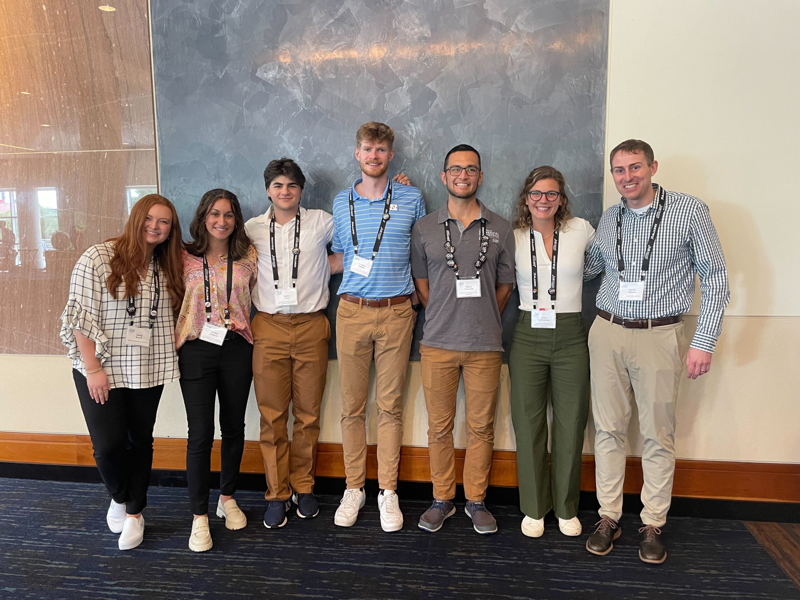
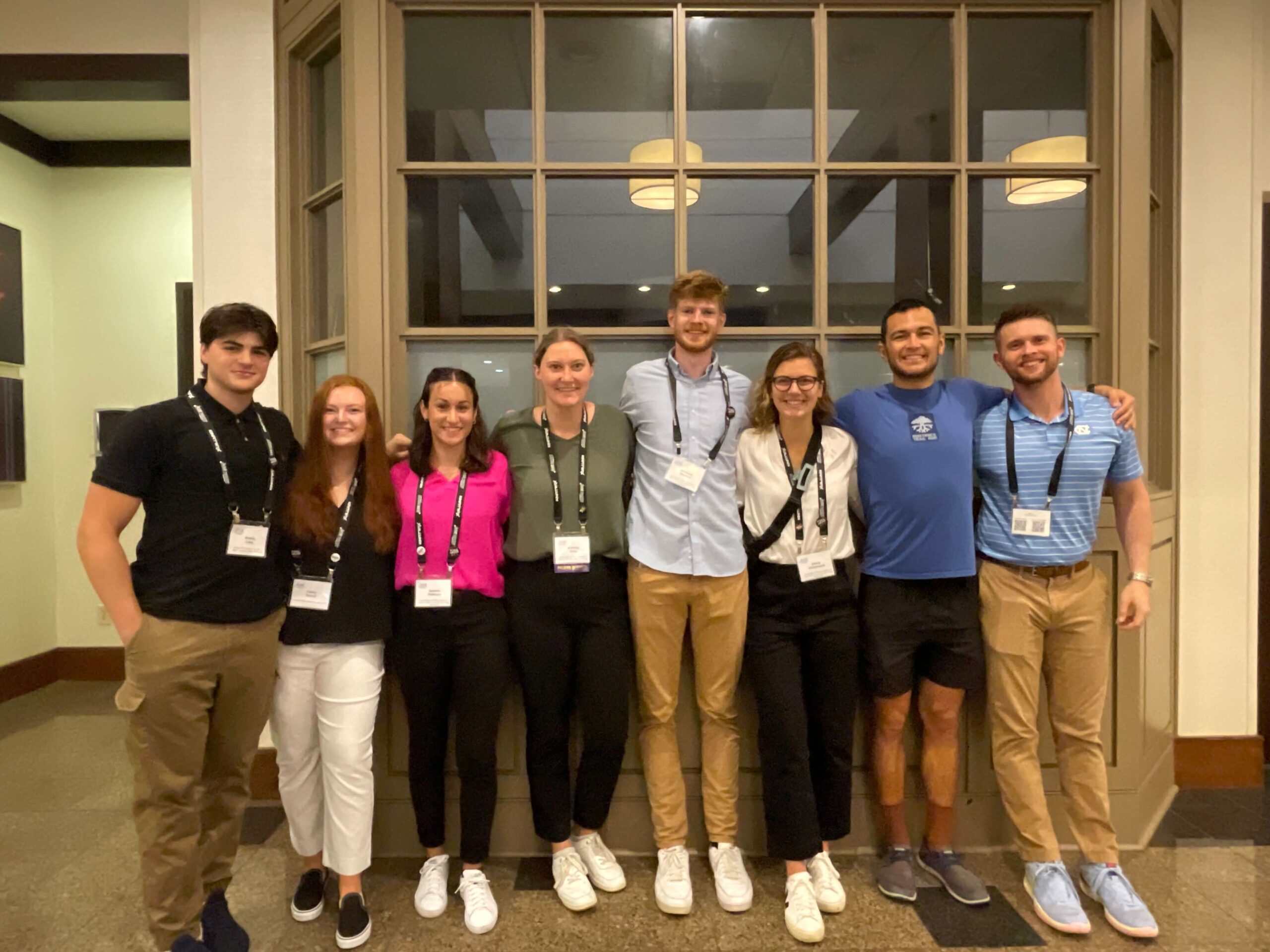
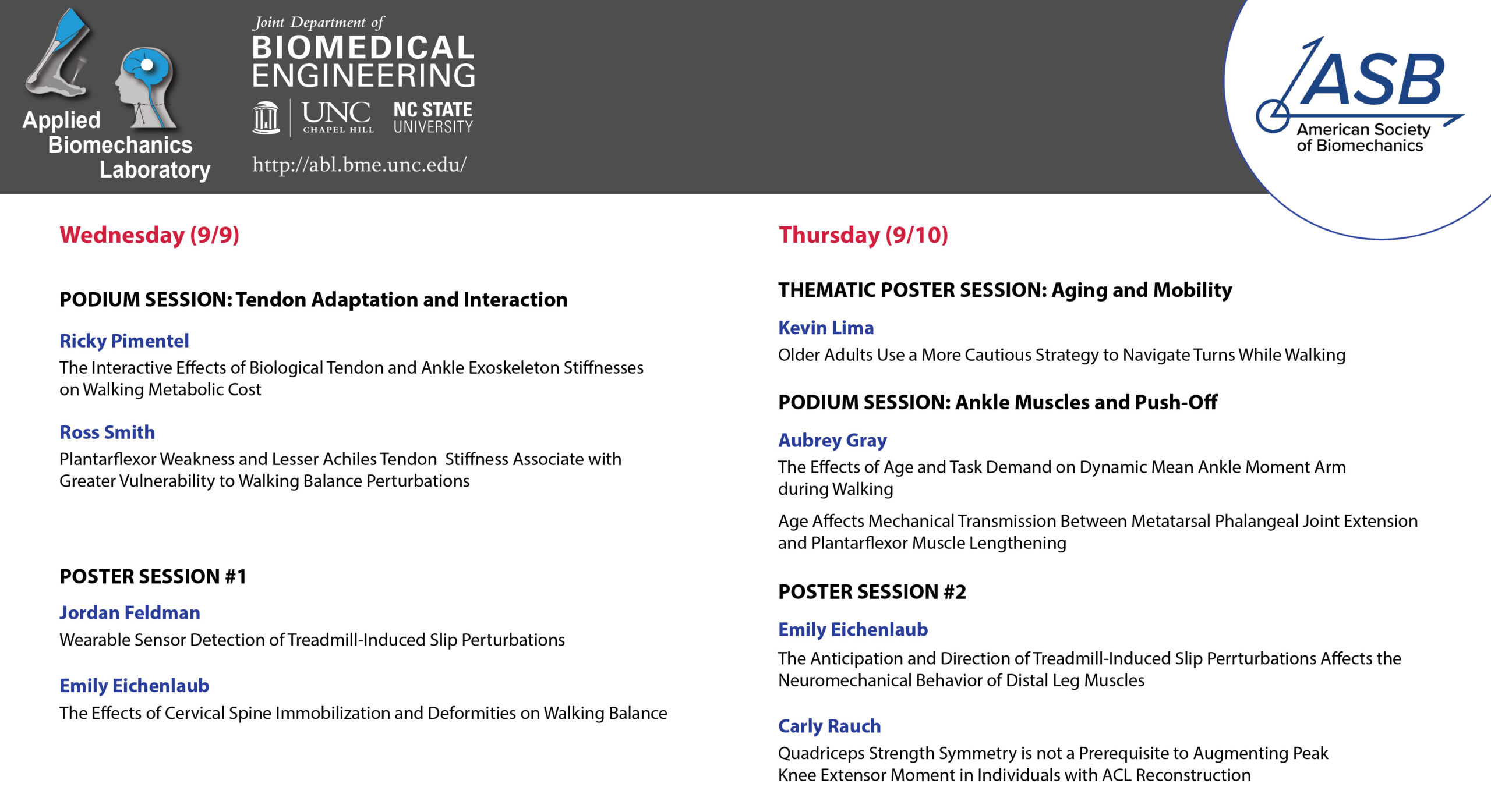
ABL Attends ASB2023

August 18, 2023

Congratulations to all our lab members that attended and presented at the 2023 Annual Meeting of the American Society of Biomechanics!


Congratulations, Emily Eichenlaub!

August 14, 2023

People | The Applied Biomechanics LabHistory of the NIH Logo | National Institutes of Health (NIH)Emily Eichenlaub, a third-year BME Ph.D. student, has received a National Research Service Award (NIH F31) from the National Institute of Aging. The award will fund her project titled “The Proactive and Reactive Neuromechanics of Instability in Aging and Dementia with Lewy Bodies.” The research will establish the effects of age and dementia on proactive and reactive neuromechanics underlying vulnerability to balance challenges. Emily will be sponsored by Dr. Jason Franz, Associate Professor in BME, and a mentoring committee that spans Engineering, Physical Therapy, Neurology and Biostatistics. Her research in the BME Applied Biomechanics Lab will pave the way for clinical translation in prescription of personalized interventions, wearable sensor monitoring to mitigate falls, and development of assistive devices with onboard monitoring of muscle neuromechanics to deliver assistance in the face of a balance challenge. Congratulations, Emily!!

 

Paper Accepted (June 2023)

June 23, 2023

Journal of Orthopaedic Research® (@JOrthopRes) / TwitterCone SG, Kim H, Thelen DG, Franz JR. 3D characterization of the triple-bundle Achilles tendon from in vivo high-field MRI. Journal of Orthopaedic Research.

The Achilles tendon consists of three subtendons that transmit force from the triceps surae muscles to the calcaneus. Potentially meaningful individual differences have been identified in subtendon morphology, with potential implications in triceps surae mechanics and function. Recently, advances in high-field magnetic resonance imaging (MRI) have previously been used to delineate boundaries within multi-bundle tendons and ligaments, including those between bundles of the anterior cruciate ligament. The objective of this study was to use high-field MRI (7T) to image and reconstruct Achilles subtendons arising from the triceps surae muscles. We imaged the dominant lower leg of a cohort of healthy human subjects (n=10) using a tuned musculoskeletal sequence (double echo steady state sequence, 0.4mm isotropic voxels). We then characterized the cross-sectional area and orientation of each subtendon between the MTJ and calcaneal insertion. Image collection and segmentation was repeated to assess repeatability. Subtendon morphometry varied across subjects, with average subtendon areas of 23.5±8.9 mm2 for the medial gastrocnemius, 25.4±8.9 mm2 for the lateral gastrocnemius, and 13.7±5.9 mm2 for the soleus subtendons. Repeatable subject-specific variations in size and position of each subtendon were identified over two visits, expanding on prior knowledge that high variability exists in Achilles tendon morphology across subjects.

Congratulations, Ricky Pimentel!

May 9, 2023

We are thrilled to congratulate Ricky Pimentel for successfully defending his PhD dissertation, titled “An Energy Audit of Human Gait: Experimental and computational assessment of how structure and function shape walking economy”. We also thank the wonderful support of his committee members, Helen Huang (UNC/NCSU BME), Mike Lewek (UNC PT), Kate Saul (NCSU MAE), and Nitin Sharma(UNC/NCSU BME). Fantastic job, Ricky! We will miss you!